Manual therapy can be performed for many reasons. One goal is often general wellness or relaxation. Another might be to address a specific pathologic myofascial condition. When massage is done for the latter, there are many terms used to describe it, including medical, therapeutic, rehabilitative, remedial, clinical, and orthopedic massage. I prefer the term clinical orthopedic manual therapy (COMT), using the word manual instead of massage to recognize and encompass all manual therapy treatment techniques a therapist can employ—massage, as well as stretching and joint mobilization.
Stocksy.
There are two major factors that usually define COMT work. One is that instead of being applied broadly across the body, the modalities are targeted at the specific structure or structures that are the underlying causative mechanism for the pain and/or dysfunction the client is experiencing. The other is the depth of the work, in other words, the force of the pressure that is being generated into the client’s tissues. For this article, I would like to explore how the depth of COMT is applied. In effect, what does it mean to generate the force needed to challenge tissue tension and find the optimal therapy zone?
Light Vs. Deep Pressure
COMT massage is generally performed with deeper pressure. Deep pressure is not always indicated or preferable to lighter work, but it’s often the key to remedying the cause of the client’s condition. So, the question might be: How do we determine when deeper work is needed? The quick answer is: when the condition is a mechanical one that is located in deeper tissues. In these cases, lighter work simply can’t reach and address the underlying cause.
Muscle Tone and Fascial Adhesions
With nearly every myofascial condition, unhealthy baseline muscle tone is involved. Most often, the resting baseline tone is overly contracted. This musculature can be described as “hypertonic,” “overly facilitated,” “locked short,” “locked long,” or, in layman’s terms, “tight.” The other factor that is usually involved, especially with more chronic conditions, is fascial adhesions. Between tight musculature and fascial adhesions, perhaps the best way to describe this is tissue densification.
Tissue Densification
Tissue densification can be divided into two categories based on the size of the area affected: a broad global area versus a focal localized area. Global densification occurs when the entire muscle, or a large portion of it, is tight. Contrasting this, focal localized densification can be caused by a myofascial trigger point (TrP) and/or fascial adhesions.
Global Tightness/Densification
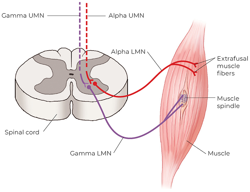
The underlying cause of global muscle tightness is not a mechanical issue in the tissue itself; rather, it lies in the nervous system’s control of baseline muscle tone. Baseline muscle tone is controlled by muscle spindle activity, which is itself controlled subconsciously by gamma motor neurons, ultimately in the brain (Image 1). With this type of tightness, even though manual treatment might be applied to the local myofascial tissue where the client is experiencing discomfort, it is ultimately directed at influencing the central nervous system.
Image 1. Muscle spindle control of baseline muscle tone. (LMN = lower motor neuron; UMN = upper motor neuron.) Image courtesy Dr. Joe Muscolino, Kinesiology, the Skeletal System and Muscle Function, 4th edition (2023), Elsevier.
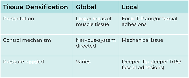
When working on a client with global tightness, depth of pressure does not necessarily need to be deep. Rather, it can vary greatly from light to deep based on the depth of work that would best convince the client’s subconscious nervous system to down-regulate their muscle tone, and this varies from client to client, as well as from one area of their body to another (see Tissue Densification Table).
Local Densification
Local densification can involve two mechanisms—myofascial TrPs and fascial adhesions. Unlike global tightness, whose root cause is in the nervous system, both TrPs and fascial adhesions are local mechanical issues situated in the myofascial tissue itself; they are not directly mediated/regulated by the central nervous system. Therefore, for manual therapy to affect these local issues, it must reach them. This means that if the target tissue is deep, the pressure must be deep. Working lightly in the area can’t cause the mechanical change needed to help resolve the condition. Let’s explore myofascial TrPs and fascial adhesions, and what role depth-of-pressure massage must play in improving each of these conditions.
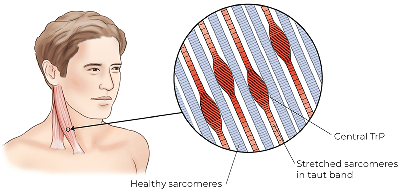
Myofascial Trigger Points:A myofascial TrP is a focal area of hypertonicity within myofascial tissue, most often within muscular tissue (Image 2).
Image 2. Myofascial trigger points. Image courtesy Dr. Joe Muscolino, The Muscle and Bone Palpation Manual, 3rd edition (2023), Elsevier.
The proposed and generally accepted primary causative mechanism is termed the energy crisis hypothesis. This posits that a lack of energy (energy crisis) is responsible for the formation and perpetuation of the TrP. To understand this, we need to understand the structure of muscle tissue.
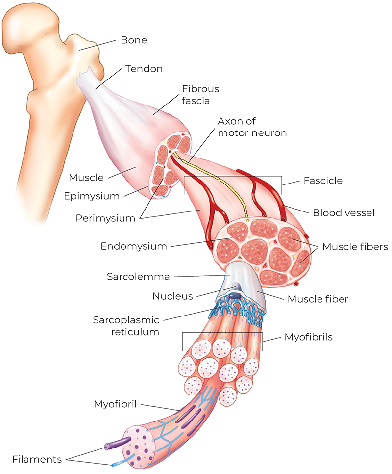
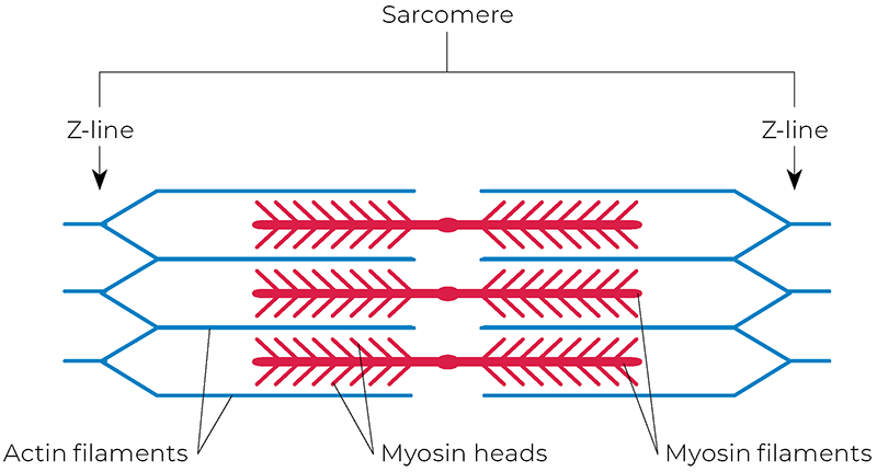
Muscle tissue structure—A muscle is composed of many muscle cells (also known as muscle fibers). Each muscle cell has myofibrils running longitudinally, and these myofibrils are composed of sarcomeres laid side-by-side and end-to-end (for context, there are approximately 10,000 sarcomeres in a linear inch) (Images 3, 4).Image 3. Muscle tissue anatomy. A muscle is composed of muscle fibers, which in turn are composed of myofibrils, which in turn are composed of sarcomeres, containing actin and myosin filaments. Image courtesy Dr. Joe Muscolino, Kinesiology, the Skeletal System and Muscle Function, 4th edition (2023), Elsevier.Image 4. Sarcomere structure. In a 2D drawing, we see two actin filaments arranged on each side around a myosin filament. Image courtesy Dr. Joe Muscolino, Kinesiology, the Skeletal System and Muscle Function, 4th edition (2023), Elsevier.
The essence of muscle contraction lies in sarcomere contraction. If we understand how a sarcomere functions, we can extrapolate to the entire muscle (the word sarcomere literally means “unit of flesh”, i.e., muscle: sarco means “flesh”; mere means “unit”). So, to make the case that a deeper TrP requires deeper pressure, let’s now delve into sarcomere structure and then apply it to the sliding filament mechanism of muscle contraction. I realize we will be getting into the weeds here, but the goal is not to teach this content; rather, it’s to refresh what was hopefully learned in massage school.
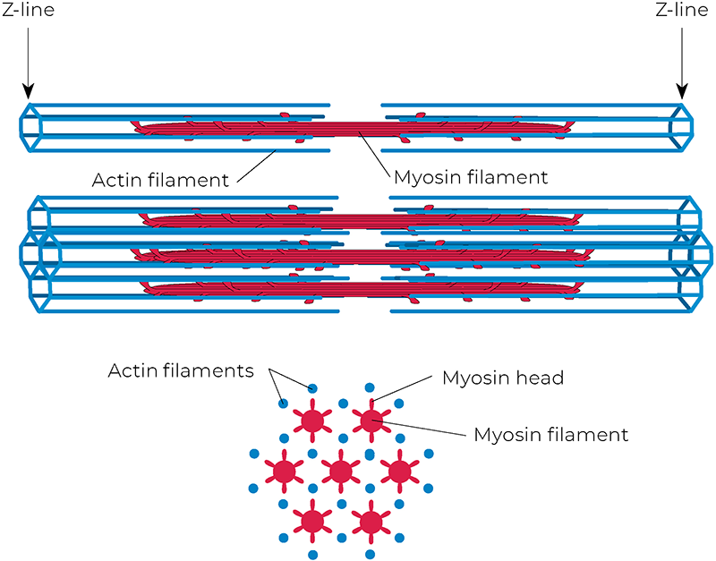
Sarcomere structure—A sarcomere is composed of a thick myosin filament in the center, with thinner actin filaments arranged around the filament on each end. In a 2D illustration, we see two actins on each side (Image 4); but in 3D, there are actually six on each side (Image 5). Image 5. The 3D sarcomere structure. There are six actin filaments arranged on each side around the myosin filament. Image courtesy Dr. Joe Muscolino, Kinesiology, the Skeletal System and Muscle Function, 4th edition (2023), Elsevier.
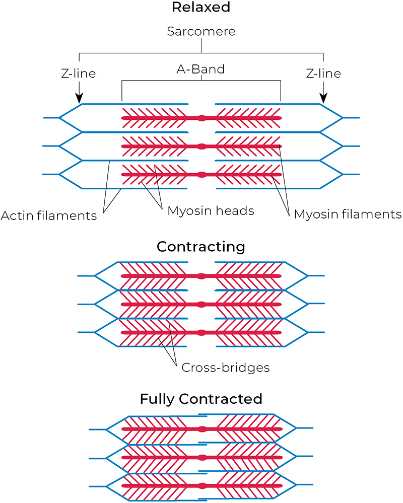
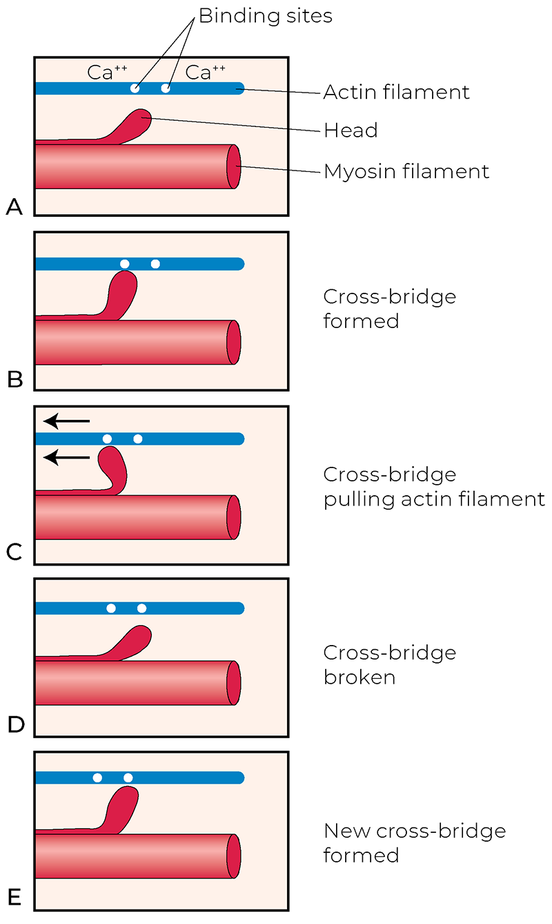
Sliding filament mechanism—It’s important to note that the myosin filament has heads that project outward toward the actin filaments. These heads can attach to the actin filaments at what are termed the actin’s active sites. When these active sites are exposed, the myosin heads grab on, forming cross-bridges, which are pre-cocked to pull the actin filaments toward the center of the sarcomere. If this pulling force is sufficiently strong, the ends of the sarcomere (Z-lines) come closer together, and the sarcomere shortens (Image 6). This mechanism is called the sliding filament mechanism because the filaments slide along each other. Given that whatever happens at one sarcomere happens at all of them, and throughout all the myofibrils and muscle fibers in the motor unit (the all-or-none response law), we can extrapolate sarcomere contraction to contraction of the muscle itself. If sarcomeres contract and shorten, the muscle contracts and shortens, yielding a concentric contraction.1Image 6. Sarcomere sliding filament mechanism. In a 2D drawing, we see two actin filaments arranged on each side around a myosin filament. Image courtesy Dr. Joe Muscolino, Kinesiology, the Skeletal System and Muscle Function, 4th edition (2023), Elsevier.
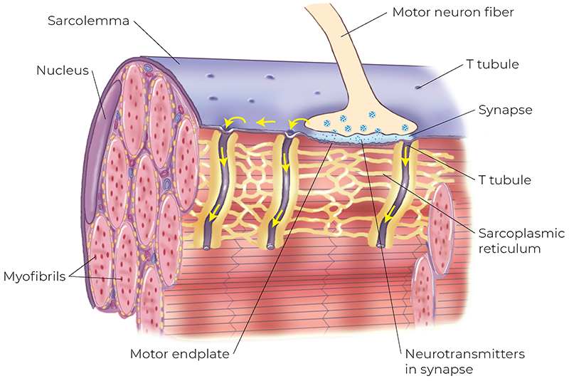
Sliding filament mechanism engagement—So, how is the sliding filament mechanism engaged? When a muscle contraction is desired, a (lower alpha) motor neuron sends a signal into the muscle fiber, causing it to release calcium that is stored in the sarcoplasmic (endoplasmic) reticulum into the fluid sarcoplasm (cytoplasm) of the muscle fiber (Image 7). The released calcium binds to the active sites of the actin filaments, causing them to be exposed. The myosin heads attach to them, forming cross-bridges, which then pull the actin filaments toward the center. What now needs to be added to the equation is that when a cross-bridge has pulled as far as it can, it must release, then grab the next active site, again pulling the actin closer toward the center, and repeat this process until the sarcomere has contracted and shortened as much as it can (Image 8).2 Image 7. A lower motor neuron innervates a muscle fiber. Image courtesy Dr. Joe Muscolino, The Muscle and Bone Palpation Manual, 3rd edition (2023), Elsevier.Image 8. Myosin-actin cross-bridge formation. Image courtesy Dr. Joe Muscolino, The Muscle and Bone Palpation Manual, 3rd edition (2023), Elsevier.
So where does the energy (and energy crisis) enter this equation? The energy for this mechanism comes from adenosine triphosphate (ATP) molecules, formed in the mitochondria by the burning of glucose in the presence of oxygen. The ATP is required during two steps in the mechanism. It is necessary to reabsorb the calcium back into the sarcoplasmic reticulum; if the calcium is not reabsorbed, then the active sites will forever remain exposed, and the myosin heads will forever stay attached, perpetuating contraction. The second place that ATP is needed is to release the myosin-actin cross-bridge; therefore, without the ATP energy, the muscle contraction cannot release. This might seem counterintuitive, but it takes energy to release a muscle contraction; a good example of this is rigor mortis (see “Rigor Mortis”).
Rigor Mortis
Shortly after death, the sarcoplasmic reticulum membrane of the muscle cell breaks down, flooding the sarcoplasm with calcium, exposing all the actins’ active sites and causing cross-bridge contraction throughout the muscle tissue, in effect locking the person into rigid contraction in whatever position the person is in at that moment. Without active ATP formation, the myosin-actin cross-bridges cannot be released. This rigor mortis (rigidity of death) contraction remains until the actual myosin-actin cross-bridges degenerate and waste away. Hence, there is no active physiology, no active generation of energy production in the muscle cell, yet the body remains in a state of contraction. Thus, we can see that once a muscle contraction begins, it can be self-perpetuating . . . it requires energy to stop the contraction. Rigor mortis helps to explain how the energy crisis hypothesis of TrP formation/perpetuation occurs.
The point of this discussion is to explain that if a muscle loses its ability to create ATP, it has a crisis of energy and a TrP forms. Once formed, it self-perpetuates. Why might muscle tissue lose its ability to generate ATP in the first place? The most likely reason is ischemia, a loss of arterial blood supply to the muscle tissue. This most likely occurs due to prolonged contraction in the muscle tissue, which hardens/densifies the tissue, thereby compressing the arteries in the area and cutting off their ability to deliver the oxygen and glucose needed by the mitochondria to create the ATP. Once the densification of the TrP occurs, it creates a vicious cycle: It cuts off its own arterial supply (ischemia), which then creates the condition wherein the TrP contraction forms, which perpetuates the ischemia, which perpetuates the TrP, etc.
Trigger Point Massage Therapy:Although there is no universal agreement on how massage therapy treatment helps treat a mysofascial TrP, and most causes of a myofascial condition are multifactorial, I believe the principal mechanism involved is promoting blood flow to the ischemic tissue of the TrP. This is essentially a mechanical problem, so it requires a mechanical solution (although neural facilitation via the nervous system can contribute, so the central nervous system can be an additional factor). The mechanical solution is generating pressure into the tissue to, in essence, milk arterial blood supply into the tissue. And now, to bring this full circle, if the TrP is located deep in the tissue, the pressure must be deep to reach it!3
Challenging Tissue-Tension Mechanical Barrier
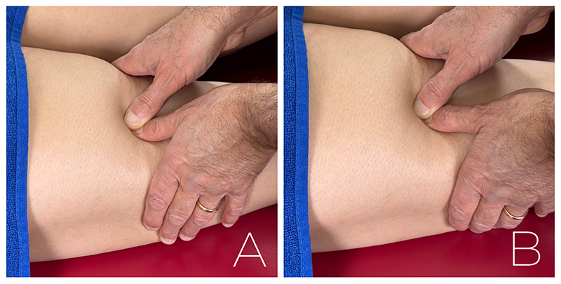
We are now at the crux of this article . . . the concept of challenging the tissue-tension mechanical barrier of densified tissue. When we first generate massage pressure into the client’s myofascial tissue, the tissue yields to our pressure, and we sink in unimpeded. But as we continue to slowly and gradually increase our pressure (Note: slowly and gradually!), we start to feel the tissue resist (Image 9).
Image 9. Reaching tissue tension with massage into the hamstrings. 9A: No resistance. 9B: Tissue tension reached. Images courtesy Dr. Joe Muscolino, The Muscle and Bone Palpation Manual, 3rd edition (2023), Elsevier.
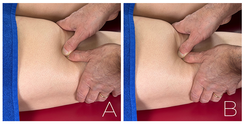
This is the point where the tissue has yielded as much as it can without us causing a change in its state. It’s at this tissue-tension barrier that we can now begin to effect a change and be therapeutic. So, for COMT to be effective, we must be able to feel this tissue-tension barrier. Next is our ability to challenge the barrier. This requires us to increase our pressure (Image 10). How much? Well, the more you challenge, the more effective the treatment is, until you reach a point where your increased pressure can cause harm.
Image 10. Challenging tissue tension with massage into the hamstrings. 10A: Tissue tension reached. 10B: Challenging tissue tension. Images courtesy Dr. Joe Muscolino.
Optimal Therapy Zone
Working in this zone of challenging tissue tension effectively but not excessively is learning how to work within what is called the optimal therapy zone. To do this, we must constantly be present and mindful. This is a kinesthetic palpatory skill, so although it does help to intellectually understand the mechanism involved (it’s amazing how the hands follow what the mind envisions), this is a skill that’s refined with physical practice and experience. So, until we have confidence in our ability to modulate our force for the optimal pressure to challenge tissue tension, it’s always safer to stay on the relatively lighter side.
Having said this, if we continually work lighter to be safer, we will not become maximally efficient at creating the healing change needed when addressing deeper tissues. So, please endeavor to learn how to deliver deep pressure comfortably (for you and the client), safely, and effectively.
Fascial Adhesions
Similar to a TrP, fascial adhesions are a local mechanical state of the tissue, not nervous-system mediated/controlled (Image 11).
Image 11A.Image 11B. Fascial adhesions. 11A: Healthy tissue. 11B: The same tissue with fascial adhesions. Reprinted with permission from Connective Tissue Research.4
Fascial adhesion densification can occur for a number of reasons, including lack of motion or injury to the tissue. Regardless of the cause, once fascial adhesions have formed, they can restrict motion in the area, causing hypomobile tissue dysfunction. To treat fascial adhesions, they must be mechanically worked with massage pressure. Whether we describe this as “melting the adhesions”or “breaking up the pattern of the adhesions,” the pressure we generate must reach the target tissue to be effective. Therefore, if the adhesions are deep in the tissue, the pressure must be commensurately deep to reach them.
Challenging Tissue Tension and Stretching
Stretching might be an even clearer example of how COMT work must challenge tissue tension to be effective. When the range of motion (ROM) at a joint is decreased, we need to stretch the client by challenging the joint to move further to increase the joint’s ROM.
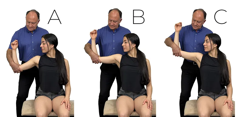
Let’s illustrate this idea with the following example: A client comes in with restricted ROM, say abduction of their arm at the glenohumeral joint is limited to 90 degrees. To increase their ROM, we need to stretch them by asking their arm to move beyond 90 degrees of abduction. During this stretch, as we move their arm up to 90 degrees, there is no resistance. At 90 degrees, we have reached tissue tension and start to feel that their tissue will resist any further movement. However, if we now stop at 90 degrees, we don’t ask for a change; consequently, we don’t improve their function because we don’t increase their ROM (we maintain the ROM, but we don’t increase it).
To be therapeutic, we need to challenge the tissue-tension mechanical barrier by moving them past 90 degrees, perhaps somewhere between 91 and 95 degrees of abduction (Image 12). We challenge tissue tension, mindful of staying within the optimal therapy zone.
Image 12. Stretching the arm into abduction at the glenohumeral joint. 12A: Below 90 degrees, there is no tissue tension. 12B: Tissue tension reached at 90 degrees. 12C: Challenging tissue tension by stretching beyond 90 degrees. Images courtesy Dr. Joe Muscolino.
Necessary Skills
I like to say that to be an excellent COMT practitioner, we need only three skill sets. The first is that we need to know anatomy. The second is that we need to be able to critically reason to figure out how to effectively treat the client (knowing anatomy and being able to think critically allow us to figure out physiology, pathophysiology, assessment, etc.). And the third skill is to be able to feel tissue-tension barriers in our client’s tissues. Being able to feel and challenge tissue-tension barriers allows us to comfortably, safely, and effectively deliver the hands-on treatment techniques needed to help treat our clients.
Notes
If the sarcomeres contract but lengthen, the muscle contracts eccentrically; and if the sarcomeres contract and stay the same length, the muscle isometrically contracts.
This description describes a concentric contraction. With an eccentric contraction, the myosin heads release and then reattach onto active sites that are closer to the center of the sarcomere, allowing the sarcomere to lengthen. With an isometric contraction, the myosin heads release and then reattach onto the same active sites, so the sarcomere stays the same length. The point here is that sarcomere contraction, whether it is concentric, eccentric, or isometric, involves cross-bridge formation and can be directly correlated to the overall muscle contraction.
There is some controversy regarding which type of stroke best facilitates increasing blood flow to the TrP. I believe that deep stroking massage is the best method for this, superior to ischemic/sustained compression. Mechanically, it makes sense—but that’s fodder for another article.
A Deeper Dive—Joint Mobilization and Tissue Tension
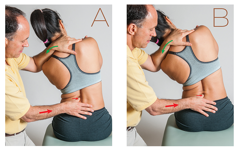
Grade IV slow oscillation joint mobilization is legal and ethical for massage therapists in most states.1 Essentially, it’s a form of specific pin-and-stretch with the target tissue usually being the intrinsic fascial (ligamentous/joint capsular) tissue of the joint.
A: Bringing the lumbar spinal joint to tissue tension. B: Challenging tissue tension with a slow oscillation stretch/mobilization force. Images courtesy Dr. Joe Muscolino.
As a stretch, we would employ the same biomechanics as we do with regular stretching (regular stretching is technically defined as “Grade III joint mobilization”). We bring the client to the end of their ROM (i.e., their tissue-tension barrier). We then ask for a little more motion by challenging that barrier. And, as in massage and stretching, we are mindful to stay within the optimal therapy zone.
Note
To be sure Grade IV slow oscillation joint mobilization is legal and ethical to perform, please check with your state regulatory board. Grade IV mobilization never involves a fast thrust; fast-thrust mobilization is defined as Grade V and is employed by chiropractic and osteopathic physicians and is never legal/ethical for massage therapists.
“Energy” or “life force” can have many definitions. As we treat clients, we should consider how its various meanings can be interpreted and applied to our work.